Monthly Archives: September 2016

eLifeの論文掲載料が2500ドルに

これまで無料だったeLifeの論文掲載料ですが。

For the first few years, it will be free to publish in eLife; we will have no publication fees or other charges to authors. (eLife All that buzz Tuesday, April 24, 2012)

2017年1月1日以降に投稿された分からは、2500ドルの掲載費用が課せられるとのことです。

This month, eLife is announcing that a fee for publication of $2,500 (USD) will take effect on January 1, 2017. All papers submitted from January 1 will be subject to the fee if they are accepted for publication, although authors with insufficient funding will be eligible for a fee waiver. (Inside eLife: Setting a fee for publication Thursday, September 29, 2016)

上記の記事では、2500ドルという設定の妥当性についても詳細に説明しています。

参考

  1. Inside eLife: Setting a fee for publication (elife news Thursday, September 29, 2016)
  2. All that buzz (elife news Tuesday, April 24, 2012)
  3. 科学出版の本当のコスト 低コストのオープンアクセスジャーナルの出現で、論文掲載料に対する出版社の付加価値に関して疑問の声が上がっている。(Nature 495, 426-429 (2013年3月28日号) | doi:10.1038/495426a Richard Van Noorden)

スペースXのマスク氏が火星植民計画を発表

メキシコ西部の都市グアダラハラ(Guadalajara)で開催中の第67回国際宇宙会議(IAC)で、スペースXのCEOイーロン・マスク(Elon Musk)氏が火星植民計画を発表しました。

Making Humans a Multiplanetary Species

参考

  1. 67th International Astronautical Congress (IAC) September 26th-30th 2016, Guadalajara, Mexico
  2. 火星コロニー構想、スペースXのマスク氏発表 「楽しい旅」に(YAHOO!JAPANニュース AFP=時事 9月28日(水)9時43分配信):”【AFP=時事】米宇宙開発企業スペースX(SpaceX)のイーロン・マスク(Elon Musk)最高経営責任者(CEO)は27日、火星に人類のためのコロニーを建設する構想を発表した。”
  3. イーロン・マスク(ウィキペディア)

匿名A氏のリスト:類似画像を含む論文 111報

当ウェブサイトは以前、「日本の科学を考えるガチ議論」の記事『捏造問題にもっと怒りを』のコメント欄に匿名A氏が書き込んだ84報を紹介しました。このコメント欄に、類似画像を含む論文等総計111報がリストアップされていたので転載します。新たな論文を指摘するものではなく、匿名A氏がこれまでに2chや『捏造問題にもっと怒りを』のコメント欄で指摘してきた論文の総まとめだそうです。

#1 Nature. 1998 Jan 1;391(6662):96-9.
Fig 1a. Lane 1 is similar to Lane 5. Lane 6 is similar to Lane 10.
#2 J Biol Chem. 2000 Mar 17;275(11):8091-6.
Fig 3A. Lane 3 is similar to Lane 8, 9, and 10. Fig 3B. Lane 1 is similar to Lane 2.
#3 Arch Biochem Biophys. 2001 Apr 1;388(1):91-9.
Fig 2A. Lanes 2-3 are similar to lanes 9-10. Fig 4B. Lanes 2-3 are similar to lanes 9-10.
#4 Diabetes. 2002 Oct;51(10):2915-21.
Fig 5. 18S of B is similar to that of C (horizontal flip).
#5 Nat Med. 2002 Jul;8(7):731-7. Epub 2002 Jun 17.
Fig 1b. +/- is partially similar to -/-. Fig 2. 18S of a is similar to that of b. Fig 5a. 28s of TNF-alpha(-) and Adiponectin(+) is similar to that of TNF-alpha(+) and Adiponectin(+).
#6 J Biol Chem. 2003 Jun 13;278(24):21344-51. Epub 2003 Apr 1.
Fig 5. Lane 2 is partially similar to lane3.
#7 Mol Cell Biol. 1996 Jun;16(6):3074-84.
Fig 7C. Lane a is similar to Lane b.
#8 J Biol Chem. 2001 Nov 2;276(44):41245-54. Epub 2001 Aug 31.
Fig 4G. PIPs are similar to those of Fig. 5d in another paper (J Clin Invest. 2001 Oct;108(7):1001-13. #9 in this list.) The date of submission of this paper is later than that of #9.
#9 J Clin Invest. 2001 Oct;108(7):1001-13.
Fig 6b. The CD36 band in the lane HF is similar to the UCP2 band in the lane HF+BADGE (horizontal flip). The CD36 band in the lane HF+BADGE is similar to the UCP2 band in the lane HF+HX531 (horizontal flip).
#10 Nat Genet. 2002 Feb;30(2):221-6. Epub 2002 Jan 30.
Fig 6. 28S in a (WAT) is similar to that of in d (BAT).
#11 Biochem Biophys Res Commun. 2004 Oct 8;323(1):242-8.
Fig 2A. The control lanes are similar to the salicylate lanes. Fig 3B. p-Akt in Lane 3 is similar to that in Lane 7. p-Akt in Lane 5 is similar to that in Lane 6. Akt in Lane 4 is similar to that in Lane 6. Fig 4. Lane 5 is similar to Lane 7 (horizontal flip).
#12 J Biol Chem. 2001 Jul 20;276(29):27519-26. Epub 2001 May 24.
Fig 6. E is similar to f. Fig 9. D is similar to e and f (enlarge).
#13 Exp Cell Res. 2002 Jan 1;272(1):23-31.
Fig7. Bone marrow cells of LZP is similar to those of CRP.
#14 Oncogene. 2002 Jan 24;21(5):844-8.
Fig 1b. These figures (HUVEC and ST2 cells) are similar to those of the COS7 cells in another paper (Fig. 6 in J Biol Chem. 2001 Jul 20;276(29):27519-26. #12 in this list.)
#15 Biochem Biophys Res Commun. 2002 Apr 26;293(1):332-7.
Fig 1. The mice of 2 weeks are similar to those of 3 weeks (vertically enlarge).
#16 J Virol. 1999 Nov;73(11):9237-46.
Fig 5B. Some bands seem to be pasted in the figures. For example, lane 3 in the left SeV/mSF figure.
#17 J Virol. 2000 Jun;74(12):5619-28.
Fig 2A. In the upper figure, 4C(-) 20 is simiar to 4C(-) 26. Fig 2B. GAPDHs of Wt 14, Wt 38, 4C(-) 14, and 4C(-) 20 are similar.
#18 J Virol. 2001 Apr;75(8):3802-10.
Fig 4C. Y1+ is similar to Y2+.
#19 J Virol. 2002 Jul;76(14):7114-24.
Fig 4B. Y2.5+ is similar to Y3+.
#20 J Virol. 2004 Jul;78(14):7443-54.
Fig 5. STAT2 of None is similar to that of Cm5.
#21 J Virol. 2007 Apr;81(7):3264-71. Epub 2007 Jan 10.
Fig 4. In the most upper figure, Sev Wt 0 is similar to Sev Wt 6 in both 2fTGH STAT1(+/+) cells and U3A STAT1(-/-) cells.
#22 Biochem Biophys Res Commun. 2002 Aug 9;296(1):194-200.
Fig 3A. Lane 1 is similar to Lane2 for GluSyn.
#23 Biochem Biophys Res Commun. 2001 Nov 30;289(2):531-8.
Fig 1 and Fig 2. 18S rRNA of Lane 2 (monocytes) in Fig 1 is similar to that of Lane 2 (alpha-GalCer-imDCs) in Fig 2.
#24 Circ Res. 2004 Jun 11;94(11):1492-9. Epub 2004 Apr 29.
Fig 2 and Fig 3. E1A in the lanes 1-2 of Fig 2A is similar to that in the lanes 2-3 of Fig 3C.
#25 J Biol Chem. 2002 Apr 5;277(14):12351-8. Epub 2002 Jan 22.
Fig 1B. D is similar to g. Fig 3B. The right part of Myc-MST1 WT is similar to that of Flag-MST1 444P.
#26 J Biol Chem. 1999 Apr 23;274(17):11995-2000.
Fig 4. EDTA is similar to Fuc.
#27 J Biol Chem. 2000 Jun 9;275(23):17233-6.
Fig 2B. Input of 0-45 is similar to that of 90-180. Fig 4. ECT2-N1(-) 45 is simialr to ECT2-N1(+) 45.
#28 J Biol Chem. 2002 Dec 27;277(52):50966-72. Epub 2002 Oct 21.
Fig 2B and Fig 4C. The actin in Fig 2B is similar to that of Fig 4C (horizontally flip.) Fig 4C and Fig 5D. The six COX bands in Fig 5D is similar to six bands of actin in Fig 4C.
#29 J Biol Chem. 2001 Mar 23;276(12):9460-7. Epub 2000 Dec 19.
Fig 1B. In the lower figure, RET-2B is similar to RET-2B/LAR.
#30 J Biol Chem. 1999 Dec 31;274(53):38251-9.
Fig 2A. 37 degrees Celsius is partially similar to 30 degrees Celsius.
#31 Nucleic Acids Res. 2000 Mar 15;28(6):1355-64.
Fig 7A. 18S rRNA of placenta is similar to that of mammary gland in another paper (Fig 2A in Mol Biol Cell. 1999 May;10(5):1637-52.)
#32 DNA Repair (Amst). 2007 Jun 1;6(6):760-9. Epub 2007 Feb 5.
Fig 5A. GAPDH of W in 5 weeks is similar to that of SP in 16 weeks.
#33 J Biol Chem. 2000 Aug 18;275(33):25146-54.
Fig 6D. pMAPK of S10A is similar to that of WT-DMSO (horizontally flip). You can pay attention to the noise of the rim.
#34 J Biol Chem. 2002 Apr 26;277(17):14355-8. Epub 2002 Mar 11.
Fig 1B. Tubulin in cytoplasm is similar to that in whole cell.
#35 EMBO J. 2002 Dec 2;21(23):6312-20.
Fig 2C. p47phox is similar to p67phox.
#36 J Biol Chem. 2003 Jul 4;278(27):25234-46. Epub 2003 Apr 25.
Fig 3A. The two lower left cells are similar between wt and P156Q.
#37 J Biol Chem. 2003 Jun 20;278(25):22908-17. Epub 2003 Apr 7.
Fig 2. MRP11-116/MRP2 is similar to MRP11-1480/MRP2.
#38 J Virol. 1999 Oct;73(10):7981-7.
Fig 1A. Lane 1 is similar to Lane 3 and 6. Lane 2 is similar to Lane 8. Lane 4 is similar to Lane 7.
#39 J Biol Chem. 2002 Jan 18;277(3):2132-7. Epub 2001 Oct 22.
Fig 2b. The FLAG band of GST-WT is similar to the GST band of WT-WT. Fig 3A. The left upper figure is similar to the left lower figure (horizontally flip).
#40 J Biol Chem. 2004 Jun 11;279(24):25474-82. Epub 2004 Mar 22.
Fig 5A. Lanes 8-9 are similar to lanes 12-13.
#41 Diabetes. 2003 Nov;52(11):2657-65.
Fig 3B. APS bands in GFP lanes seem to be pasted in. IR beta bands in GFP lanes are similar to those in APS(YF) lanes.
#42 J Biol Chem. 1999 Nov 5;274(45):32309-17.
Fig 4A. Lane 1 is similar to Lane 15 (horizontally flip). Lanes 12-13 are similar to Lane 16-17 (horizontally flip).
#43 J Biol Chem. 2000 Sep 1;275(35):26856-63.
Fig 9C. Mock-transfected cell (-) is somewhat similar to Mutant probe (-). Mock-transfected cell oligo TRE is somewhat similar to Mutant probe Ang II.
#44 J Biol Chem. 2000 Feb 11;275(6):4369-73.
Fig 3. GAPDHs of Time 4, 5, and 6 are similar in PAO+. Fig 4. iNOS mRNA of Lane +-+- is similar to that of Lane +–+.
#45 Hepatology. 2000 Nov;32(5):1037-44.
Fig 3. 3h None is similar to 5h Hypo.
#46 J Hepatol. 2004 Apr;40(4):616-23.
Fig 4A. Phospho-Akt of 2h(-) is similar to that of 2h(+).
#47 Am J Physiol Endocrinol Metab. 2005 May;288(5):E876-82. Epub 2004 Dec 21.
Fig 4A. 28S and 18S in lanes 1-6 are similar to those in another paper (Fig 1A in Biochem Biophys Res Commun. 2004 May 14;317(4):1075-9.)
Fig 5A. In the adiponectin bands, Lanes 1-4 are similar to lanes 12-15. Lanes 6-7 are similar to lanes 9-10.
#48 Biochem Biophys Res Commun. 2001 Apr 27;283(1):255-9.
Fig 2. V/Vsp in lanes 1-3 is similar to V/Vsp in lanes 7-9, STAT1 in lanes 10-12, and STAT1 in lanes 13-15. IRF9 in lanes 10-12 are similar to that in lanes 13-15.
#49 J Virol. 2002 Dec;76(24):12683-90.
Fig 8. In the Blot:FLAG, FL is similar to FLMT in the two middle lanes (vertically enlarge).
#50 J Biol Chem. 2003 Oct 24;278(43):41654-60. Epub 2003 Aug 13.
Fig 4. HSF-1s of OSC19-MP(mock) C and IFN in Fig 4A, from total cell lysate, are similar to those of OSC19 cytosol C and IFN in Fig 4C. HSF-1s of OSC19-MP(STAT-1) C and IFN in Fig 4A, from total cell lysate, are similar to those of OSC19-MP cytosol C and IFN in Fig 4D.
#51 J Med Virol. 2006 Apr;78(4):417-24.
Fig 2. GAPDH of RSV 5 is similar to that of inactivated SARS 1. GAPDH of FluAV 1 is similar to that of inactivated SARS 2. Fig 5. GAPDH of RSV 5 is similar to that of inactivated SARS 1. GAPDH of FluAV 1 is similar to that of inactivated SARS 2.
#52 Cancer Lett. 1999 Jul 19;142(1):23-30.
Fig 2B. hTERT of lane D is similar to that of lane F2.
#53 Leukemia. 2000 Jul;14(7):1260-5.
Fig 1b and 3b. hTERT of Fig 1b is similar to that of Fig 3b (horizontally flip).
#54 Biochem Biophys Res Commun. 2004 Apr 2;316(2):528-32.
Fig 1. 15 N is partially similar to 30 N.
#55 Cancer Lett. 2008 Mar 18;261(2):226-34. Epub 2007 Dec 21.
Fig 1. K562/hTERT 1 is similar to K562/hTERT 10.
#56 Cancer Res. 2006 Oct 15;66(20):9913-20.
Fig 4C. ADAM28 of Day3 is similar to that of Day 31 (vertically enlarge). Fig 5C. Lane 1 is similar to Lane 4.
#57 Biochem Biophys Res Commun. 2005 Mar 25;328(4):1232-43.
Fig 2. Two exon 3 figures are similar.
#58 Biochem Biophys Res Commun. 2000 Aug 11;274(3):603-8.
Fig 5. Two right lower bands in Fig 5A are similar to two left lower bands in Fig 5B.
#59 Biochem Biophys Res Commun. 2001 May 11;283(3):707-14.
Fig 3C. In the lower figure, the left four bands are similar to the middle four bands and the right four bands.
#60 Nat Cell Biol. 1999 Dec;1(8):479-85.
Fig 4a. Western(PS) of A246E is partially similar to that of delta E9.
#61 J Biol Chem. 2001 Jan 19;276(3):2108-14. Epub 2000 Oct 12.
Fig 1. Lanes N and H in Fig 1C are similar to Fig 1D.
#62 J Biol Chem. 2001 Nov 16;276(46):43446-54. Epub 2001 Sep 10.
Fig 1a and Fig 2a. Ten actin bands of Fig 1a are similar to those of Fig 2a.
#63 J Biol Chem. 2002 Apr 12;277(15):12931-6. Epub 2002 Jan 25.
Fig 3b. PY20 of Src is similar to Lysate of Src.
#64 Circulation. 2002 Jun 18;105(24):2893-8.
Fig 2. ERK of Fig 2C is similar to that of Fig 2D (horizontally flip, change brightness and contrast).
#65 J Biol Chem. 2002 Mar 8;277(10):8076-82. Epub 2002 Jan 4.
Fig 3. Cyclin D1 and actin of 694F are somewhat different with those of delta p85.
#66 J Biol Chem. 2005 Feb 11;280(6):4929-39. Epub 2004 Nov 24.
Fig 2A. Bcl-2 and actin of ED(-) are similar to those of ED(+).
#67 J Biol Chem. 2005 Apr 1;280(13):13163-70. Epub 2005 Jan 25.
Fig 5E. The left four lanes of CHO-B are similar to the right four lanes of CHO-B.
#68 J Biol Chem. 2001 Mar 30;276(13):9688-98. Epub 2000 Dec 14.
Fig 2C. Lane +— is similar to Lane +-+-. Fig 5B. Lane 1 is similar to Lane 3.
#69 J Biol Chem. 2001 Dec 14;276(50):47642-9. Epub 2001 Oct 10.
Fig 1B. Lane 2 is similar to Lane 4 (horizontally flip). Fig 5A. ERKs of lanes 1-4 are similar to those of lanes 5-8 (horizontally flip). Fig 7A. The upper two bands of pSG5 are similar to those of ER beta (horizontally flip).
#70 J Biol Chem. 2001 Feb 2;276(5):3459-67. Epub 2000 Oct 23.
Fig 7B. pSG5 is similar to ER beta (horizontally flip).
#71 J Biol Chem. 2002 Sep 6;277(36):33490-500. Epub 2002 Jun 26.
Fig 1B. ERKs of lanes 1-3 are similar to those of lanes 4-6. Fig 3A. Lanes 1-2 of Caov-3 are similar to lanes 3-4 of Caov-3 (horizontally flip). Fig 4A. Lanes 1-2 of BAD are similar to lanes 3-4 of BAD. Fig 6B. Phospho-Raf of Lane 2 is similar to that of Lane 5.
#72 Endocrinology. 2004 Jan;145(1):49-58. Epub 2003 Sep 18.
Fig 3B. Akt of lanes 1-2 is similar to that of lanes 3-4.
#73 Clin Cancer Res. 2004 Nov 15;10(22):7645-54.
Fig 1D. Akt of lanes 1-2 is similar to that of lanes 3-4.
#74 Endocrinology. 2004 Mar;145(3):1302-13. Epub 2003 Nov 26.
Fig 8B. Lane 1 of A2780 is similar to Lane 3 of Caov-3.
#75 J Biol Chem. 2004 May 28;279(22):23477-85. Epub 2004 Mar 16.
Fig 2A. Lanes 2-4 of actin are similar to lanes 6-8 of actin (horizontally flip).
#76 J Biol Chem. 2000 Nov 10;275(45):35051-62.
Fig 7C. Lanes i, j and k of abDbf4p are somewhat similar.
#77 Proc Natl Acad Sci U S A. 2000 Dec 5;97(25):13824-9.
Fig 4A. The most upper figure of insulin is similar to that of IGF-1.
#78 J Biol Chem. 1999 Mar 26;274(13):8531-8.
Fig 5C. In the Ad5IkB lane, Bcl-2 is similar to Bcl-x (horizontally flip and vertically enlarge). In the Ad5LacZ+TNF lane, Bcl-2 is partially similar to Bcl-x (vertically enlarge).
#79 FASEB J. 2001 May;15(7):1218-20.
Fig 1A. Akt of Cont is similar to that of VEGF.
#80 Nat Med. 2001 Mar;7(3):317-23.
Fig 2a. Actin of Astrocytes is similar to that of another paper (Fig 5C in J Biol Chem. 2001 Feb 2;276(5):3046-53. Epub 2000 Oct 20.)
#81 J Biol Chem. 2003 Jan 17;278(3):2058-65. Epub 2002 Nov 7.
Fig 2. wt is similar to delta alpha 1.
#82 J Biol Chem. 2001 Sep 7;276(36):34259-69. Epub 2001 Jul 2.
Fig 7. SRE-352 is similar to SRE-344 (vertically enlarge).
#83 J Biol Chem. 2005 Mar 18;280(11):10468-77. Epub 2005 Jan 7.
Fig 5B. IB:anti-V5 of STAM1 is similar to that of STAM1-mUIM.
#84 Cancer Res. 2007 Jun 1;67(11):5162-71.
Fig 2A. The well of WT is similar to that of control.
#85 Nature. 2008 Jul 17;454(7202):345-9.
Fig. 1c. The beta-actin of the lane 1 is similar to that of the lane 2.
Fig. 3b. The beta-actin of the lane 1 is similar to that of the lane 2.
Fig. 3e. The alpha-MHCs of the lanes 1, 4, and 6 and the GATA-4 of the
lane 5 are similar.
Fig. 3e. The GATA-4 of the lane 1 is similar to that of the lane 6.
Supplementary Fig. S3e. The bands of Nkx2.5 (-) and GATA4 BP4-2 are similar.
Supplementary Fig. S3f. The bands of Nkx2.5 (-) and beta-actin (-) are
similar (resized).
#86 J Biol Chem. 2004 Mar 19;279(12):11384-91.
Fig. 3. The MLC-2v is similar to the cardiac alpha-actin.
#87 Nat Med. 2009 Sep;15(9):1082-7.
The CT scan of WT (HS/HS) of Supplemental Fig. 2c is similar to those
of Pre operation of Supplemental Fig. 2d, G4 (HS/HS) of Supplemental
Fig. 2c, and G4 Trp53+/-(HS/HS) of Supplemental Fig. 2c.
The bars of G4 (HS/HS) and G4 Trp53+/- (HS/HS) for lean tissue/BW in
the right of Supplemental Fig. 2c are identical for mean and SEM.
#88 Nat Cell Biol. 2004 Jun;6(6):499-506.
The lower right band in the Fig. 2f is similar to the right band of Fig. 2h.
The upper band in the lane 3 in Fig. 2e is similar to the band in the
lane 2 in Fig. 3e.
#89 Biochem Biophys Res Commun. 2004 Sep 10;322(1):310-9.
Fig. 2b. The 28Ss of the lane 1-3 are similar to those of the lane 4-6.
#90 J Biol Chem. 1999 Mar 19;274(12):8231-9.
TTF1 of Fig. 2b is similar to NKE2 of Fig. 2c and CSX+ GATA-4+ of Fig. 7.
#91 Biochem Biophys Res Commun. 2000 Apr 21;270(3):1074-9.
Fig. 2a. GAPDH of WT At is similar to that of TG At. GAPDHs of WT Vt,
TG Vt, and TG Sk are similar.
#92 Circulation. 1999 Nov 16;100(20):2100-7.
Fig. 3b. DM 10 min in GST-c-Jun (1-79) is similar to DM 60 min
(horizontally flip.)
#93 Circulation. 2003 Dec 16;108(24):3024-30.
Fig. 5c. The upper band in Sham TG is similar to that of I/R TG.
#94 Circulation. 1998 May 19;97(19):1952-9.
Fig. 4. 18S of the right of Wild Type Banding is similar to that of
AT1a KO Banding.
18S of the left of Wild Type Sham is similar to that of Wild Type Banding.
18S of the right of Wild Type Sham is similar to those of the both
lanes of AT1a KO and that of the left lane of ShamAT1a KO Banding.
#95 Circ Res. 1999 Mar 5;84(4):458-66.
Fig. 2. The (-) in the upper MBP is similar to D.N.Cdc42.
#96 EMBO J. 2000;19(20):5533-41.
Fig. 3a. The upper band of +/- is similar to the band of -/-. Pay
attention to that the three “+” over the figure are not identical.
Fig. 7b. The eight error bars in the right panel are similar.
#97 Hypertension. 1998 Jan;31(1):50-6.
Fig. 5. The two lanes of S(SHRSP) in 24 wk are similar.
#98 Circ Res. 1998 Oct 5;83(7):752-60.
Fig. 1c. The 5 min is similar to the 3h (horizontally flip.)
#99 J Am Soc Nephrol. 2003 Mar;14(3):584-92.
Fig. 1c. The three lanes of ERK are similar to those of p38.
#100 Hypertens Res. 2005;28(5):447-55.
Fig. 2b. Con is similar to ALD/Spi.
#101 J Pharmacol Sci. 2005;98(4):372-9.
Fig. 4. BNP is similar to MCP-1.
#102 Circulation. 2003;107(10):1411-7.
Fig. 5. The lanes 1 and 2 of alpha-tubulin are similar to the lanes 3 and 4.
#103 Hypertension. 2006; 48:628-636.
All of the pictures and graphs of Fig. 1 HT are similar to those of
Fig. 5 HT-SD for.
All of the pictures and graphs of Fig. 2 HT is similar to Fig. 7 HT-SD
for except for the ICAM Western.
The ICAM Western in Fig. 2 NT is similar to that of Fig. 7 HT-SD
(horizontally flip.)
The actin of NT in Fig. 2 is similar to that of HT-Chimera in Fig. 7.
#104 Mol Pharmacol. 2005;67(5):1666-73.
Fig. 2A p-Src is similar to Fig. 2A Ser-STAT3 and Fig. 2C Tyr-STAT3.
The left 7 lanes of Fig. 2A Src are similar to the right 7 lanes of Fig. 2C Src.
The right 7 lanes of Fig. 2A ERK are similar to the left 7 lanes of Fig. 6A ERK.
The right 7 lanes of Fig. 2A Jak2 are similar to the left 7 lanes of
Fig. 2C Jak2.
Fig. 2C p-Pyk2 is similar to Fig. 2C Pyk2.
Fig. 2C Ser-STAT1 is similar to Fig. 2C Ser-STAT3.
The right 5 lanes of Fig. 2C Ser-STAT1 are similar to the right 5
lanes of Fig. 4A siRNA-MKP-1 MKP-1 (horizontally flip.)
The left 2 lanes of Fig. 4A siRNA-Lamin A/C alpha-SM actin are similar
to the left 2 lanes of Fig. 4A siRNA-MKP-1 alpha-SM actin.
The rightest lane of Fig. 4A siRNA-Lamin A/C alpha-SM actin is similar
to that of Fig. 4A siRNA-MKP-1 alpha-SM actin.
The left 7 lanes of Fig. 4B siRNA-Lamin A/C ERK are similar to the
right 7 lanes of Fig. 4B siRNA-MKP-1 ERK.
Fig. 6A Pyk2 is similar to Fig. 6C Pyk2.
Fig. 6A Src is similar to Fig. 6A Jak2.
Fig. 6A p-ERK is similar to Fig. 6C p-ERK.
The left 4 lanes of Fig. 6A p-Tyk2 are similar to the left 4 lanes of
Fig. 6A Tyr-STAT1 and the left 4 lanes of Fig. 6C Tyr-STAT3.
The right 4 lanes of Fig. 6A Tyr-STAT1 are similar to the right 4
lanes of Fig. 6A STAT3.
The left 7 lanes of Fig. 6A Tyr-STAT3 are similar to the left 7 lanes
of Fig. 6C Tyr-STAT1.
The right 4 lanes of Fig. 6A Tyr-STAT3 are similar to the left 4 lanes
of Fig. 6A STAT3 (horizontally flip.)
Fig. 6C p-Src is similar to Fig. 6C p-Tyk2 and Fig. 6C Ser-STAT1.
Fig. 6C Src is similar to Fig. 6C Tyk2 and Fig. 6C STAT1.
Fig. 2C Src is similar to Fig. 2C STAT3 (horizontally flip.)
#105 Arterioscler Thromb Vasc. 2012 Jun;32(6):1453-9.
Fig. 5. oxLDL (-) of A is similar to oxLDL (50ug/ml) of B.
#106 Nature. 1998 Jul 2;394(6688):92-6.
Fig. 2b. -TGF-beta, M-2[Evi-1(-)] is similar to +TGF-beta, E-5[Evi-1(+)].
#107 Circulation. 2006 Aug 29;114(9):953-60.
Supplemental Fig. IV. Something wrong in DKO+Vehicle of ICAM-1.
#108 Circulation. 2002 Sep 10;106(11):1397-402.
Fig. 3. 2-c is similar to 3-c (rotate.) The experimental condition for
3-c is different between the main text and the figure legend.
#109 Circulation. 2002 Mar 12;105(10):1240-6.
The lanes 1-3 in Skeletal alpha-actin in Fig. 3a is similar to the
lanes 2-4 of Skeletal alpha-actin in Fig. 1B.
#110 Arthritis Rheum. 2000 Feb;43(2):259-69.
There are a lot of duplications in the actin of Fig. 3B.
#111 J Clin Invest. 1999; 104(2):137-146
Fig. 2a. Src. Control is similar to AxCATcsk(moi) 100 (horizontally flip.)
Fig. 4b. IL-6. Control is similar to AxCASLacZ. Actin. Control is
partially similar to AxCATcsk(moi) 100.
Fig. 6d. Csk. Ax1w1 is similar to AxCATcsk Day42.

参考

  1. 新参者のために私がこれまで2ちゃんねるやこのコメント欄でしてきた111報の指摘を再び紹介します。匿名Aとは何か? この指摘をした人物のことです。あとはどーでもいい低脳な話をしているだけ。全て新規指摘ではないので騒がないでください。騒いだらか恥ずかしいです。新しく科学部に配属されたマスコミの方々は、一つ一つ丁寧に調べ、私が隠した数百万人の命に関わるリスクを見つけてください。(日本の科学を考えるガチ議論 捏造問題にもっと怒りを コメント欄 匿名A)
  2. 酷似する画像を含む生命科学論文がインターネット上で大量に指摘される

DNAの解析でキリンは4種に分類される

動物のキリンは、種としてはひとつだけから成っており、亜種としていくつかに分かれるとこれまで考えられてきました。しかし、見た目は同じようにみえるキリンですが、DNAレベルでの解析から4種に分けるべきだと主張する論文が、カレント・バイオロジーに掲載されました。DNAの差がこれほど大きければ、これらの4種間が野生で交尾することはないだろうと著者は述べています。

“We have studied the genetic relationships of all giraffe subspecies from across the continent. We found, that there are not only one, but at least four genetically highly distinct groups of giraffe, which apparently do not mate with each other in the wild. This we found looking at multiple nuclear genes considered to be representative of the entire genome” says Professor Axel Janke, researcher at the Senckenberg Biodiversity and Climate Research and Professor at the Goethe University in Frankfurt, Germany. “Consequently, giraffe should be recognized as four distinct species despite their similar appearance.” (EurekAlert! 8-Sep-2016)

参考

  1. Multi-locus Analyses Reveal Four Giraffe Species Instead of One (Current Biology Published: September 8, 2016)
  2. Giraffes more speciose than expected: Gene analyses reveal that there are not one, but 4 giraffe species (EurekAlert! Goethe University Frankfurt Public Release: 8-Sep-2016)
  3. キリン、実は4種類? 独チームが遺伝子解析で新説(朝日新聞DIGITAL ワシントン=小林哲 2016年9月10日11時28分):”キリンを遺伝子解析した結果をもとに、アフリカ全体でただ一つの種に属するとしてきた従来の学説に反して「異なる四つの種に分類されるべきだ」とする新説を、ドイツなどの研究チームが米専門誌カレント・バイオロジー電子版に発表した。”
  4. キリン、単独種でなく4種 (ロイター 2016年 09月 9日 02:05 JST):”これまで亜種を含めて一つの種と考えられていたキリンが、実は独立した四つの種に分かれているとする遺伝子解析の結果を、ナミビアの「キリン保護基金」やドイツの研究者らのチームが8日付の米科学誌カレントバイオロジーに発表した。”
  5. キリンは4種、遺伝子解析=保護策に活用も-独大学など(NIFTY NEWS/時事通信 2016年09月09日 14時14分):”これまでキリンは1種とされてきたが、ドイツ・フランクフルト大などは遺伝子解析で4種に分かれると発表した。アフリカに生息するの遺伝子を解析すると、四つの種に分類されることが分かった。体の形などに基づく従来の分類では一つの種で、種の下の亜種レベルで九つに分類されることが多い。”
  6. キリン 単独種ではなく4種に分けられると研究で明らかに(LIVEDOOR NEWS 2016年9月9日 13時1分):”米科学誌カレント・バイオロジー(Current Biology)に掲載された論文によると、研究チームはアフリカ全域のキリン190頭の皮膚からサンプルを採取し、そのDNAを調べた。”
  7. birth of a giraffe (キリンの雄同士の儀式化した闘争行動、雄と雌の交尾、出産の映像)
  8. Giraffe Vs Giraffe Deadliest Fight Ever Seen – Nat Geo Wild (オス同士の激しい闘争行動。後頭部に打撃を与えて、相手を気絶させるキリン)
  9. 数秒以内に終了するキリンの交尾

  10. 9割が同性愛! キリンの異常な愛情とは? (世界中の仰天ニュースをお届け! 2014年1月18日):”戦いを通じて雄同士で愛情が芽生えるのでしょうか。何と争った後には雄同士で首を愛撫しあい、そのままセックスを始めるというのです。驚くべきはその割合。動物学者の推定によるとおよそ74%~95%ものキリンが雄同士でセックスをするのだとか。”


3D女子高生 Saya テルユカプロジェクト

2016年10月5日追記

シーテックジャパン(Combined Exhibition of Advanced Technologies (CEATEC)JAPAN)2016年10月4日-10月7日 幕張メッセで、動くSayaさんが発表されました。ちなみに動きに関しては、実際に人間にモデルとなってもらい、その体の動きのデータを取り込んで用いています。
CEATEC JAPAN 2016 シャープブース CG美少女「Saya」動画デモ

参考

  1. 動く「Saya」に会える! 「CEATEC JAPAN 2016」のシャープブースで、女子高生CGキャラ「Saya」の8K動画を公開(SHARP Blog 2016年10月4日):”10月4日から7日まで幕張メッセで開催している「CEATEC JAPAN 2016」の当社ブースにて、これまで静止画での作品発表だった女子高生CGキャラ「Saya」の8K動画を、当社の8K映像モニターで公開しています。”


* * *

TELYUKA(テルユカ)プロジェクトの3D女子高生 Sayaさんは、3Dコンピュータグラフィックスなんだそうですが、どうみてみ実写にしか見えません。

saya2016small

(画像:TELYUKA(テルユカ)プロジェクトのウェブサイト telyuka.com より)

参考

  1. telyuka.com
  2. 3DCG女子高生Saya2016がヤバい出来に。完全に不気味の谷を超えた(世永玲生)(engadget日本版 BY 世永玲生(Reo Yonaga) 2016年09月09日 18時15分):”フリーランスの夫婦CGユニット”TELYUKA”(テルユカ)のプロジェクト「Saya」の2016バージョンがつい先ほど公開されました。”
  3. CG女子高生「Saya」が超リアル 「不気味の谷」超えた執念の手描き (withnews.jp 信原 一貴 2016年05月17日):”昨年秋、一人の美少女「Saya」の画像がツイッターに投稿され、大きな反響を呼びました。実在の少女を写したような圧倒的にリアルな画像が、すべてコンピューターグラフィックス(CG)で描かれたものだったからです。あれから半年、Sayaの進化は止まっていません。”
  4. 超リアルな女子高生CG『Saya』の作者が実践する、フォトリアルキャラクター制作術|CGWORLD 2015 クリエイティブカンファレンス個別レポ(6)(CGWORLD.jp 2016.01.16 SAT):”2015年11月22日(日)、文京学院大学 本郷キャンパスにて催された「CGWORLD 2015 クリエイティブカンファレンス」。本稿では、世間一般からも大きな注目をあつめた超フォトリアルな女子高生CGキャラ『Saya』を生み出したTELYUKAの2人によるセッション「フォトリアルキャラクターメイキング」をレポートする。 “
  5. “かぎりなく実写”な女子高生CGキャラ『Saya』で国内外から注目をあつめるデジタルアーティスト、「TELYUKA」(テルユカ)とは、何者?(CG・映像の専門情報サイト CGWORLD.jp スペシャルインタビュー 2015.11.19)

データ捏造で研究費2億ドル 損害賠償請求訴訟

2016年9月1日のサイエンス誌のニュース記事によると、アメリカのデューク大学らの研究者は不正な研究データに基づいて総額2億ドルの研究費助成金を国から得ていたとして、研究者および大学は国に与えた損害額の3倍を支払うよう訴えられていることが明らかになりました。

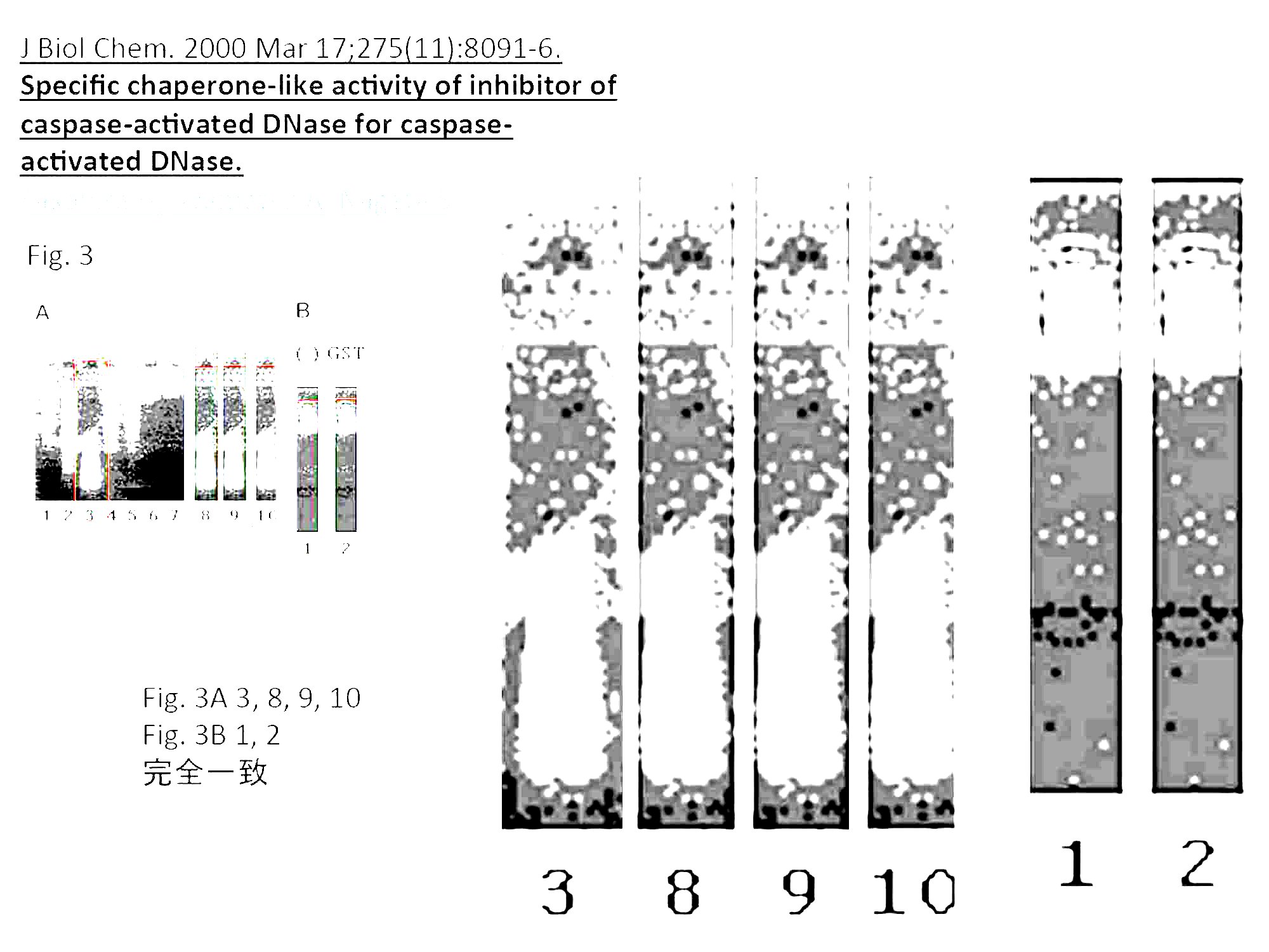
デューク大学のWilliam Foster教授の研究室で働いていたErin Potts-Kant博士が研究データの捏造などの不正を行い、Potts-Kant博士とFoster教授の共著で論文を出していましたが、結果的に、彼女が関与した15報の論文が現在までに撤回されていす。Potts-Kant博士およびFoster教授は不正論文に基づいて研究費を獲得しさらに不正論文を生産することを長期わたって繰り返した結果、不正に得た研究資金の総額が2億ドル(助成件数60件以上)にも上っていました。データ捏造に直接関与したPotts-Kant博士、不正を隠蔽したFoster教授およびデューク大学は、国に与えた損害を弁済すべしというのが、今回の訴えの内容です。

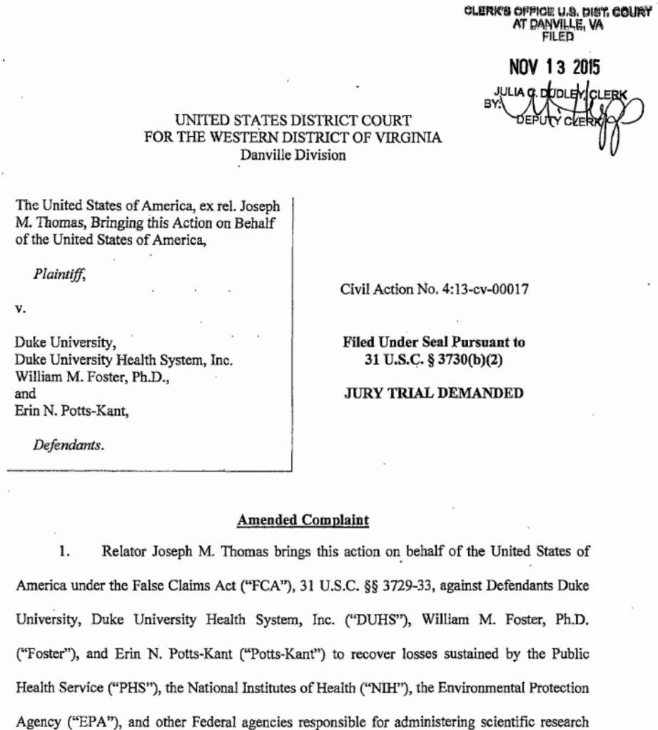
Relator Joseph M. Thomas brings this action on behalf of the United States of America under the False Claims Act (“FCA”), 31 U.S.C.§§3729-33,  against Defendants Duke University, Duke University Health System, Inc,(“DUHS”), William M.Foster, Ph.D. (“Foster”), and Erin N. Potts-Kant (“Potts-Kant”) to recover losses sustained by the Public Health Service (“PHS”), the National Institutes of Health (“NIH”), the Environmental Protection Agency (“EPA”), and other Federal agencies responsible for administrering scientific aresearch grants. (Civil Action No.4:13-cv-00017)

裁判でこの訴えが認められると、実際に支払いを求められるのは不正で獲得した研究費の金額ではなく、ペナルティが科せられます。訴状によると、デューク大学、Foster教授、Potts-Kant博士らの被告側に対して、国に与えた損害金額の3倍の金額の支払いなどが求められています。

WHEREFORE, Relator, on be half of the United Sates, prays that judgment be entered in their favor and against Defendants as follows:
1. That Defendants pay the United States triple the amount of its damages to be determined, plus civil penalties of up to $11,000 for each false claim, statement, or record; (Civil Action No.4:13-cv-00017)

告発者は、当時デューク大学に所属していた研究者で、訴えが認められた場合、インセンティブとして数百万ドル(数億円)を手にする可能性があるとのことです。今回のこの訴訟は、国のお金を横領した個人や会社などに対して、その返還を求めて、個人が国を代表して訴訟を起こす制度False Claims Actに基づくものです。

ちなみに、Potts-Kant博士は今回問題にされている研究そのものに関する不正とは別に、25000ドル以上の研究費を着服し、アマゾンやウォルマート、ターゲットなどでの買い物に使っていたかどで2013年に逮捕されています。

サイエンスの記事:

”And last month, a U.S. district court unsealed a whistleblower lawsuit filed by a former colleague of Potts-Kant. It accuses the researcher, her former supervisor, and the university of including fraudulent data in applications and reports involving more than 60 grants worth some $200 million. If successful, the suit—brought under the federal False Claims Act (FCA)—could force Duke to return to the government up to three times the amount of any ill-gotten funds, and produce a multimillion-dollar payout to the whistleblower.” (Whistleblower sues Duke, claims doctored data helped win $200 million in grants. Science News By Alison McCook, Retraction WatchSep. 1, 2016 , 2:00 PM)

訴状は公開されていて、内容を読むことが可能です。

DukeSuedCivilActionNo4-13-cv-00017

Former researcher sues Duke, alleges Uni used improper data to receive funding. Duke University The Chronicle By Staff Reports | Friday, September 2 訴状へのリンク)


参考

  1. Duke Is Wake-up Call for Research Compliance (bna.com April 4, 2018) The case is in the discovery phase in preparation for a possible trial, according to filings tracked by Bloomberg Law.
  2. Judge Refuses to Dismiss Whistleblower’s False Research Data Suit Against Duke University and Two Faculty Members (The Health Law Firm Thursday, May 11, 2017) On April 27, 2017, a federal judge in North Carolina refused to dismiss a False Claims Act (FCA) lawsuit against Duke University and some of its faculty.
  3. Survival of Duke Research Whistle-Blower Case Could Mean More Suits (bna.com May 2, 2017)  Duke University and two Duke researchers will have to face claims they defrauded the government on NIH research grants ( United States ex rel. Thomas v. Duke Univ. , M.D.N.C., No. 1:17-cv-00276-CCE-JLW, order 4/25/17 ). Former Duke University employee Joseph M. Thomas sufficiently stated claims against Duke and the researchers, Judge Catherine C. Eagles of the U.S. District Court for the Middle District of North Carolina said in an April 25 order refusing the defendants’ bid to dismiss the suit.
  4. Joseph Thomas vs. Duke University: Lawsuit Update 4.28.17 (Scribd)
  5. Former researcher sues Duke, alleges Uni used improper data to receive funding (Duke University The Chronicle By Staff Reports | Friday, September 2)
  6. Whistleblower sues Duke, claims doctored data helped win $200 million in grants. (Science News By Alison McCook, Retraction Watch Sep. 1, 2016 , 2:00 PM)
  7. Embezzlement, 15 retractions, and a whistleblower could add up to trouble for Duke (Retraction Watch Written by Ivan Oransky September 1st, 2016 at 2:00 pm)
  8. William Michael Foster, PhD Professor in Medicine Depart of Medicin Duke University School of Medicine
  9. Lawsuit: Scientific fraud at Duke cost government $200 million in grant awards (The News&Observer By Jane Stancill September 2, 2016 7:22 PM)
  10. Duke researcher adds another retraction in JCI, bringing count to 15 (Retraction Watch Written by Alison McCook May 11th, 2016 at 12:05 pm):”We’ve found another retraction for Erin Potts-Kant, a former researcher at Duke, bringing her total to 15.”
  11. So you want to be a whistleblower? A lawyer explains the process (Retraction Watch Written by Alison McCook March 18th, 2015 at 9:30 am)
  12. False Claims Act (Wikipedia): The False Claims Act (31 U.S.C. §§ 3729–3733, also called the “Lincoln Law”) is an American federal law that imposes liability on persons and companies (typically federal contractors) who defraud governmental programs. It is the federal Government’s primary litigation tool in combating fraud against the Government.[1] The law includes a qui tam provision that allows people who are not affiliated with the government, called “relators” under the law, to file actions on behalf of the government (informally called “whistleblowing” especially when the relator is employed by the organization accused in the suit). Persons filing under the Act stand to receive a portion (usually about 15–25 percent) of any recovered damages. As of 2012, over 70 percent of all federal Government FCA actions were initiated by whistleblowers.

2008年ノーベル化学賞Roger Y. Tsien博士が死去

2008年ノーベル化学賞受賞のカリフォルニア大学サンディエゴ校教授ロジャー・ツェン博士(64)が、訪れていたオレゴン州ユージンで2013年8月24日に亡くなられました。

UC San Diego Chancellor Pradeep Khosla said that Tsien apparently died while on a bike trail, the San Diego Union-Tribune reported (http://bit.ly/2bSZn8Z), but the cause of death had not been determined.(abcnews.go.com)

参考

  1. UCSD Nobel laureate Roger Tsien dies (The San Diego Union-Tribune By Gary Robbins | 10:27 a.m. Aug. 31, 2016 | Updated, 6:51 p.m.)
  2. Nobel Laureate Roger Tsien Dies, Age 64(UC San Diego News Center August 31, 2016 | By Scott LaFee)
  3. Nobel Prize Winner Roger Tsien Dies; Helped Track Cells (ABC NEWS By The Associated Press SAN DIEGO — Aug 31, 2016, 7:16 PM ET)